Table of Contents
A new wave of brain-computer interface research in China is exploring sound-based alternatives to implanted electrodes. Startups aim to use acoustic energy to change neural activity without surgery. They promote focused ultrasound for noninvasive stimulation, while acknowledging technical limits and privacy concerns about decoding brain states.
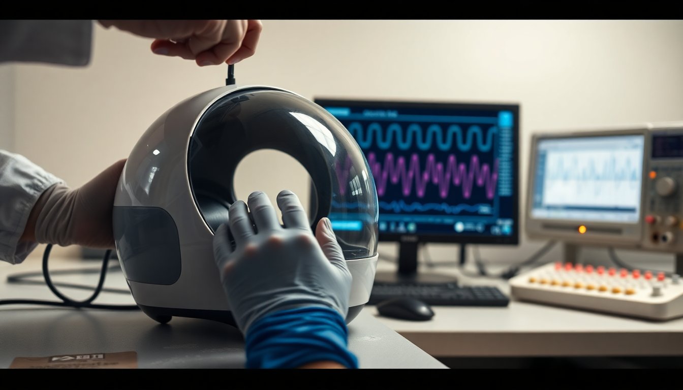
One company founded in Chengdu, with offices in Shanghai and Hong Kong, is developing systems that both stimulate and, ultimately, record brain signals with high-frequency acoustic waves. The approach builds on medical ultrasound’s safety record in imaging and some therapies, but applies the same physical principle directly to neural circuits rather than only to tissue visualization.
What focused ultrasound brings to brain-computer interfaces
Building on the previous section, researchers are shifting the focus from electrodes to acoustic energy that interacts with neural tissue. Conventional implanted brain-computer interfaces record electrical activity directly from neurons. By contrast, ultrasound-based BCIs apply focused acoustic energy to target brain regions without opening the skull.
Stimulation without scalpels
Focused ultrasound can serve multiple roles depending on acoustic parameters. It can create high-resolution images, ablate pathological tissue, or modulate neural circuits with subablative exposures. Clinical use of focused ultrasound for Parkinson’s disease and certain brain tumors provides an existing therapeutic foundation for developers exploring neuromodulation.
Clinical trials and peer-reviewed preclinical studies show that low-intensity, pulsed ultrasound can alter neuronal excitability. From the patient perspective, the appeal is clear: noninvasive stimulation reduces surgical risk and shortens recovery time compared with implanted sensors. The technology also promises more flexible targeting of deep structures that are difficult to reach with surface stimulation.
Technical challenges remain. The skull attenuates and distorts ultrasound, complicating precision and dose control. Achieving millimetre-level spatial resolution without thermal damage requires careful calibration of frequency, intensity and pulse duration. Safety endpoints and standardized dosimetry must be established through rigorous clinical trials and long-term follow-up.
Regulatory and clinical uptake will depend on robust evidence from randomized trials and real-world data. If validated, ultrasound-based BCIs could expand therapeutic and interface options while lowering procedural barriers for patients. Ongoing research will determine which clinical indications and device designs meet the thresholds for safety and efficacy required by regulators and clinicians.
Researchers and companies developing noninvasive neuromodulation argue that the chief benefit is stimulation of brain targets without surgical incision. For patients, that could mean lower procedural risk compared with implanted systems. Early pilot studies have focused on brain regions tied to the emotional component of pain. Clinical trials show that targeted stimulation reduced pain intensity for days in some participants. Firms describe staged rollouts that start with clinic-based machines and may progress to supervised wearable systems for home use.
Clinical programs and product roadmaps
Developers are designing programs that pair incremental clinical evidence with device iterations. Initial studies are clinic-based to control delivery and monitor safety. As safety and efficacy data accumulate, manufacturers plan supervised use models suitable for outpatient or home settings.
According to the scientific literature, pilot trials and phase 1 programs prioritize short-term analgesic effect and tolerability. Clinical trials show that measurable pain reductions occur in subsets of patients, but responder rates and durability vary across studies. Regulatory approval will depend on randomized data that demonstrate consistent benefit beyond placebo and acceptable risk profiles.
From the patient perspective, supervised wearable paths could increase accessibility while maintaining clinician oversight. The data real-world evidence required to support home use will likely include device performance, adherence, and adverse-event monitoring. Medical ethicists note the importance of clear patient selection criteria and informed consent when treatments shift from clinic to home.
Manufacturers plan iterative product roadmaps tied to evidence milestones. Early commercial launches would target specialized clinics and pain centers. Subsequent versions would incorporate user-friendly interfaces and remote monitoring to support supervised home therapy. As emerges from clinical development, regulators and payers will assess whether the devices meet thresholds for safety, efficacy, and cost-effectiveness required for broader adoption.
Clinical trials show that the anterior cingulate cortex plays a central role in the emotional and motivational aspects of pain. According to the scientific literature, modulating this region can alter how patients perceive suffering rather than changing sensory signals alone. From the patient’s point of view, that distinction could mean relief from the distress that makes chronic pain disabling.
The developer’s first-generation system is a stationary, clinic-operated focused-ultrasound device. Future iterations are described as helmet-like units designed for supervised home treatment. Those next-generation designs aim to widen access while preserving clinician oversight and safety monitoring.
Beyond chronic pain, the company cites research avenues that include depression, stroke rehabilitation, Alzheimer’s disease and sleep disorders. Each indication engages different neural networks and will require separate evidence packages and regulatory pathways. Peer-reviewed data and phase 3 clinical trials will be needed to establish efficacy for each target.
From targeted relief to broader indications
Peer-reviewed data and phase 3 clinical trials will be needed to establish efficacy for each target. From targeted relief to broader indications, researchers are exploring whether ultrasound can not only modulate but also read brain activity.
Can ultrasound also read the brain?
Functional ultrasound imaging is emerging as a research tool that maps blood flow changes linked to neural activity. The technique detects hemodynamic signals with higher spatial resolution than many noninvasive alternatives. According to the scientific literature, these signals correlate with neuronal activation in animal models and small human studies.
Translating flow signals into direct, real-time readouts of neural firing remains technically demanding. Signal sources overlap, and skull properties attenuate and distort ultrasound waves. Controlled studies are required to separate vascular responses from genuine neural signals. Clinical trials show that rigorous validation against established methods is essential.
From the patient perspective, a noninvasive sensor that reads brain states would transform monitoring and care. Real-world data could enable closed-loop therapies that adjust stimulation based on ongoing activity. However, ethical questions about privacy, consent and data ownership must be addressed before deployment.
Parallel efforts combine ultrasound with biomarkers and machine learning to interpret complex patterns. As emerges from peer-reviewed work, multimodal approaches—pairing ultrasound with EEG or MRI—improve reliability. Regulatory pathways will hinge on demonstrated safety, reproducibility and clinical benefit.
For now, ultrasound shows promise as a window into brain function, not as a standalone neural decoder. Ongoing clinical trials and larger peer-reviewed studies will determine whether it can meet the standards required for diagnostic or monitoring use in patients. Advances in signal processing and device design are the next steps to watch.
Advances in signal processing and device design are the next steps to watch.
Researchers are exploring whether ultrasound can do more than modulate circuits. The proposed model is a closed-loop system that would detect signatures of pain, depression or other states and deliver targeted pulses in response. Compared with implanted electrodes that record local electrical activity, an ultrasound approach could survey larger brain regions with a single device.
Clinical studies show that converting acoustic or hemodynamic signals into reliable neural readouts remains difficult. Acoustic measures reflect tissue motion and pressure waves rather than direct neuronal currents. Hemodynamic proxies lag behind electrical events by seconds and have coarser spatial resolution. These factors complicate the translation of raw signals into high-resolution maps of neural activity.
From an engineering perspective, three challenges stand out. First, algorithms must distinguish neural patterns from vascular and motion noise. Second, hardware needs to maintain safe energy levels while achieving sufficient spatial specificity. Third, validation will require comparison with gold-standard recordings in peer-reviewed clinical trials and real-world datasets.
Dal punto di vista del paziente, a noninvasive system that both reads and writes brain signals could reduce surgical risks and broaden access. The data real-world evidenziano the need for rigorous, evidence-based benchmarks before clinical deployment. As device prototypes mature, independent replication and phase 3–level trials will determine whether ultrasound can reliably interpret brain states at the resolution clinicians require.
Following assessments required for phase 3–level validation, two technical barriers persist that shape feasibility and clinical timelines.
First, the human skull attenuates and distorts acoustic waves. This interference reduces targeting accuracy and degrades signal fidelity. Researchers sometimes employ implants or skull windows in laboratory studies to bypass that limitation. True noninvasive precision sufficient for clinical decision making remains unproven.
Second, most ultrasound monitoring detects hemodynamic changes such as blood flow or volume shifts. These signals lag behind the millisecond-scale electrical spikes of neurons. That temporal delay constrains use cases that require real-time decoding, including instant speech reconstruction.
Global context and ethical considerations
Clinical studies show that technological readiness alone does not determine adoption. Regulatory approval, reimbursement policy and health-system capacity also matter. According to the scientific literature, devices that perform well under controlled conditions can face new challenges in routine care.
From the patient perspective, safety and informed consent are paramount. Implant-based work raises distinct risks compared with noninvasive approaches. The ethics of risk-benefit trade-offs must be examined in prospective clinical trials with patient-centred endpoints.
The technology raises equity concerns. High-cost platforms and specialist centers could concentrate access in wealthy regions. Real-world data highlight disparities in uptake for other advanced neurotechnologies, suggesting similar patterns may appear here.
Dual-use and privacy risks require governance. Imaging or decoding techniques that infer mental states create novel vulnerabilities for misuse. Independent, peer-reviewed evaluation of algorithms and data governance frameworks will be essential.
Evidence-based pathways are needed to move from experimental demonstration to clinical integration. Robust multicentre trials, standardized outcome measures and transparent reporting should precede widespread deployment. The ultimate test will be whether these tools deliver measurable benefit for patients and health systems.
International programs are pursuing similar approaches, and high-profile investments and startup launches abroad signal growing global interest in combining neuroscience with advanced sensing and AI. Experts caution that consumer-ready products remain years from market and that progress must be validated by rigorous clinical testing. Clinical trials show that rigorous phase 3–level evaluation is essential to demonstrate safety and measurable benefit for patients and health systems.
Policy and privacy implications are equally urgent. Devices that analyze brain states would collect highly sensitive neural data, requiring clear governance on storage, access and consent when algorithms infer mood, pain or other internal states. From the patient’s perspective, consent frameworks must protect autonomy and limit secondary uses. The convergence of major tech firms and neurotechnology companies amplifies both therapeutic potential and privacy risk, making regulatory clarity and independent oversight critical for responsible development.
Focused ultrasound BCIs: promise amid technical and ethical hurdles
Focused ultrasound BCIs remain experimental but show potential to expand noninvasive brain therapies. Developers must overcome skull-related physics and reliably map hemodynamic signals to neural activity.
Clinical trials show that hemodynamic measures can correlate with neural states, yet translating those correlations into real-time control remains a technical challenge. From the patient’s perspective, a truly noninvasive interface could reduce procedural risk and broaden eligibility for neurotherapies.
Progress will depend on rigorous, peer-reviewed research, transparent regulatory frameworks, and independent oversight. Public debate about data ownership, consent, and secondary use of sensitive brain-derived information will be essential as the field moves from laboratories toward clinical settings.
The trajectory of this quieter form of brain interfacing will reflect both scientific advances and how regulators, clinicians, and patients resolve questions of safety, efficacy, and privacy.